- Metrology - Laboratory
- Analytical Instrumentation
- 3D imaging system
- Hitech Instruments
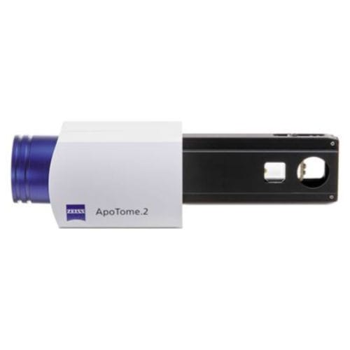
3D imaging system Apotome.2optical2D
Add to favorites
Compare this product
Characteristics
- Options
- optical, 2D, 3D
Description
Optical sectioning using structured illumination for brilliantly resolved 2D or 3D fluorescence images free from stray light - now includes Apotome deconvolution algorithm to further improve your image stacks.
Create optical sections of your fluorescent samples – free of scattered light. With structured illumination, you know that only the focal plane appears in your image: ApoTome.2 recognizes the magnification and moves the appropriate grid into the beampath. The system then calculates your optical section from three images with different grid positions without time lag. It’s a totally reliable way to prevent scattered out-of-focus light, even in your thicker specimens. Operate your system just as easy as always. You get images with high contrast in the best possible resolution – simply brilliant optical sections.
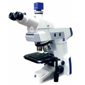
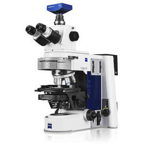
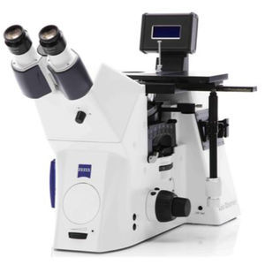
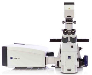
•Compatible with AxioObserver, AxioImager, AxioZoom
•Increased resolution in x,y and z
•Perfect images with all magnifications - Apotome now features automatic grid selection and setting optimization
•Compatible with all standard light sources and filter sets
•Brilliant 3D rendering, even with thick specimens
•All Apotome images can be viewed as optical sections, conventional fluorescence or raw data
•New deconvolution algorightm can optional be used to further improve image stacks
•Free Zen lite viewer includes Apotome and 3D image support
Catalogs
No catalogs are available for this product.
See all of Hitech Instruments‘s catalogs*Prices are pre-tax. They exclude delivery charges and customs duties and do not include additional charges for installation or activation options. Prices are indicative only and may vary by country, with changes to the cost of raw materials and exchange rates.


