- Metrology - Laboratory
- Laboratory Equipment
- Electron microscope
- THERMO FISHER SCIENTIFIC - MATERIALS SCIENCE
- Products
- Catalogs
- News & Trends
- Exhibitions
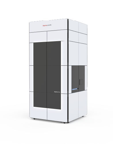
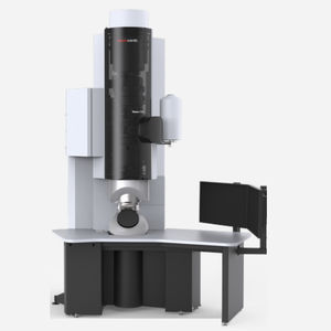
Scanning transmission electron microscope Spectra Ultrafor quality controlfor materials researchfor semiconductors
Add to favorites
Compare this product
Characteristics
- Type
- scanning transmission electron
- Technical applications
- for quality control, for materials research, for semiconductors
- Electron source
- cold field emission
Description
Scanning transmission electron microscope for imaging and spectroscopy of beam sensitive materials.
Spectra Ultra Scanning Transmission Electron Microscope
To truly optimize TEM and STEM imaging, EDX and EELS may require acquisition of different signals at different accelerating voltages. The rules may vary from sample to sample but, it is generally accepted that: 1) the best imaging is done at the highest possible accelerating voltage above which visible damage will occur, 2) EDX, especially when mapping, benefits from lower voltages with increased ionization cross-sections, thus yielding better signal-to-noise ratio maps for a given total dose, and 3) EELS works best at high voltages to avoid multiple scattering, which degrades the EELS signal with increasing sample thickness.
Unfortunately, acquisition at different accelerating voltages on the same sample without losing the region of interest—all during a single microscopy session—is not possible. At least, until now.
Imagine a Thermo Scientific Spectra 300 S/TEM:
• That can truly be operated at different voltages (all the voltages between 30 and 300 kV for which alignments were purchased) in a single microscopy session
• Where changing from an accelerating voltage to any other one takes about 5 minutes
• That can accommodate a radically different EDX concept with a 4.45 srad solid angle (4.04 srad solid angle with an analytical double tilt holder)
With the new Spectra Ultra S/TEM, the accelerating voltage becomes an adjustable parameter, just like probe current, and the massive Ultra-X EDX system enables chemical characterization of materials too beam-sensitive for conventional EDX analysis.
Catalogs
No catalogs are available for this product.
See all of THERMO FISHER SCIENTIFIC - MATERIALS SCIENCE‘s catalogsOther THERMO FISHER SCIENTIFIC - MATERIALS SCIENCE products
Transmission electron microscopes
Related Searches
- Thermo Fisher Scientific microscope
- Thermo Fisher Scientific laboratory microscope
- Inspection microscope
- Thermo Fisher Scientific benchtop microscope
- Thermo Fisher Scientific measuring microscope
- Thermo Fisher Scientific analysis microscope
- Teaching microscope
- Thermo Fisher Scientific high-resolution microscope
- Thermo Fisher Scientific industrial microscope
- Thermo Fisher Scientific automated microscope
- Thermo Fisher Scientific compact microscope
- Thermo Fisher Scientific materials research microscope
- Thermo Fisher Scientific quality control microscope
- Research microscope
- Thermo Fisher Scientific 3D microscope
- Thermo Fisher Scientific electron microscope
- Thermo Fisher Scientific motorized microscope
- Observation microscope
- High-speed microscope
- Thermo Fisher Scientific microscope for the electronics industry
*Prices are pre-tax. They exclude delivery charges and customs duties and do not include additional charges for installation or activation options. Prices are indicative only and may vary by country, with changes to the cost of raw materials and exchange rates.


